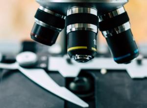
Finding the correct microscope depends on knowing which technique is required to achieve your experimental goals.
Below are brief descriptions of common microscopy techniques with experimental examples to help guide you to the correct equipment.
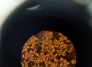
Confocal microscopy provides improved optical sectioning and resolution compared to the widefield microscopy, by eliminating or reducing the out-of-focus light.
FLIM is an imaging technique based on the measurement of a molecule’s fluorescence decay rate, or how long a fluorescent molecule remains in its excited state before decaying to its ground state
FRAP is an imaging based technique used to measure the diffusion of fluorescently labelled molecules in biological samples.

Forster Resonance Energy Transfer (FRET) offers a tool to examine the physical distance of < 10 nanometers between two molecules, which is in a range for inter-molecular interactions.
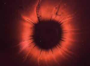
Light sheet microscopy utilizes a planar illumination technique to provide a unique way of volumetric imaging for thin (<10um) samples like cultured cells as well as for thick specimens like tis
Live cell imaging has been used extensively to provide information about the dynamic nature of cell biology.
Multiphoton microscopy, a concept first introduced by Maria Goeppert-Mayer in 1931, uses pulsed-high power far red and infrared wavelengths to excite femtoliter sized volumes in live and fixed sam
Superresolution Microscopy comprises a variety of optical/computational tools that circumvent the resolution limit defined by the diffraction of light.
In TIRF microscopy, an evanescent light wave is used to illuminate the sample in close proximity to the coverslip.